
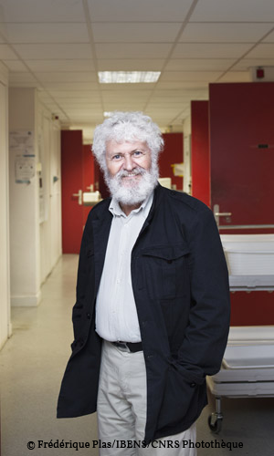
Karsenti was already riding high. Having joined the CNRS three years earlier, he was sent to San Francisco
VOYAGE INSIDE THE CELL – MITOSIS
Credits
from one division to the next and trigger mitosis,” Karsenti recounts. “We knew that the microtubules, the tiny protein tubes that form the skeleton of the cell, are able to organize themselves just before division into a structure called the mitotic spindle. By pulling on the chromosomes, which by then have been duplicated, the microtubules in the spindle separate the two sets of homologous chromosomes and position them at the two poles of the spindle, enabling the formation of two daughter cells. Back then, the scientific community thought that the centriole,
In 1985, the biologist took off for Heidelberg, where he joined the department of cell biology at the European Molecular Biology Laboratory (EMBL). There he continued his Californian research as part of a new team, with a new goal: to identify the factor(s) that cause chromosome condensation, a vital phase in mitosis. Except during the brief period of division, the cell’s DNA does not come in the form of chromosomes, usually represented as Xs and Ys. Most of the time, the DNA strands are unraveled, forming a mass at the center of the cell’s nucleus, like thousands of unwound thread reels tangled up on a huge rug. It is not possible to separate the threads into two perfectly equal bunches without rolling them up first. The same thing happens within the cell: before being divided into two batches, the chromosomes’ DNA has to be “rewound”. The question is: which protein is responsible for this tedious process?
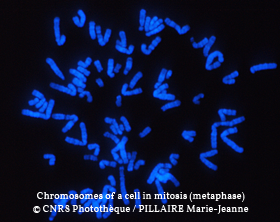
MITOSIS AND SPINDLE
Credits
MICROTUBULES
Credits
In parallel, a team led by the British genetician Paul Nurse had identified the protein pair responsible for the phenomenon: kinase cdc2, an enzyme that activates other proteins by adding phosphate, and cyclin, which activates the kinase. The latter, in turn, activates proteins responsible for the condensation of DNA into chromosomes and the assembly of the spindle. For this breakthrough, Nurse, along with Leland Hartwell and Timothy Hunt, was awarded a Nobel Prize in 2001. “ At the same time,” Karsenti comments, “Marcel Dorée had begun purifying the kinase from Xenopus (Xenopus is a genus of frogs whose eggs, produced in large quantities, are used as a biological model in the laboratory) eggs in my lab at the EMBL. He completed the project with a student in Montpellier. Personally, I think he should have been named co-winner of the Nobel.”
In Heidelberg, Karsenti and Dorée follow in Nurse’s footsteps, showing that the kinase remains inhibited until the cyclin concentration reaches a certain threshold, and that it deactivates spontaneously after ten minutes.
“Given the autoinhibition of the kinase activity,” Karsenti continues, “I started thinking about other instances of self- organization in biology. While working with my students on the mitotic spindle during that period, I realized that a phenomenon of this type was taking place: at a certain point in the cell cycle, ‘motors’ linked to the microtubules adopt a collective behavior and organize these small building blocks into a spindle. From microscopic chaos emerges an ordered, quasi-macroscopic structure. It’s fascinating!”
By the end of the 1980s, the mystery surrounding the universal molecular motor of the cell cycle was about to be solved.
THE CLOCK IN EVERY CELL
Each living cell is equipped with a clock-like mechanism that controls its cycle, as demonstrated by Eric Karsenti and his teams over years of experiments.
« Already at the University of San Francisco, I started ‘breaking’ Xenopus eggs in 1981-82, isolating the cytoplasm for in vitro experiments, » the researcher recounts. In Heidelberg, with the help of my first student, Marianne Felix (now a senior researcher at the CNRS), I used this method to examine how the kinase/cyclin complex was regulated over time.”
The two scientists noticed that the complex did not become active immediately upon entry into the cytoplasm. Only after a series of reactions would the cyclin reach the critical threshold, after gradually accumulating. This latency time is of great importance: it enables the complex to autoactivate at once, rather than gradually, inducing a “clean break” in the form of mitosis
Once activated, the complex remains active for 10 minutes. After that, the cyclin level drops sharply, before gradually building up again0for the next cycle. “We were able to demonstrate that the kinase/cyclin complex itself causes this drop,” Karsenti explains. “If cyclin alone is inserted into our ‘broken eggs’, its concentration remains constant. But if active kinase is inserted into in an egg containing cyclin, the latter disappears after 10 minutes. The kinase triggers a second clock that determines the moment of its own inactivation. In other words, the genetic products of the cell cycle are generated in the proportions needed for the self-organization of a biological clock, enabling an alternation between interphase and mitosis —in Xenopus eggs, every 30 minutes on the dot. It’s amazing!”
THE MOTORS
THAT DRIVE MITOSIS
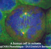 In the lab, Eric Karsenti’s team has shown that without the action of certain molecular motors, proteins that use the cell’s energy to move along the microtubules, the mitotic spindle cannot form properly.
But to understand how the protein motors, microtubules and chromosomes behave collectively during the assembly of the spindle, the biologist relied on digital simulations0performed in cooperation with physicists.
Using this tool, “We understood that during mitosis, the motors bind to the microtubules,” Karsenti reports. “By forming bridges between them, and by all moving in the same direction, toward the two poles of the cell, they rearrange the microtubules in a spindle around the chromosomes.”
These same microtubules induce the separation of the chromosomes, and grow shorter with the help of the motors, exerting a pull on the chromosomes. Biologists call this depolymerization, and liken the process to a necklace losing its beads. Thus pulled apart, the X chromosomes are cut in half lengthwise, with each half going to one of the daughter cells.
“This is another case of self-organization,” Karsenti concludes. “The collective behavior of the chromosomes, microtubules and motors induces the formation of a ‘machine’, the spindle that separates the chromosomes. We are beginning to understand how complex cellular functions emerge from what seems to be chaos.”
Meanwhile, in 1988, Eric Karsenti embarked on a new adventure. In Roscoff (Brittany), he initiated the first Jacques Monod conference on the cell cycle, with a view to fostering interdisciplinarity — a principle that would guide his entire career. Hosted by the CNRS and INSERM through the Institute of Biological Sciences (INSB) and the Institute of Ecology and Environment (INEE), the conference series continues to this day, with four to six events scheduled each year, most often in Roscoff. Discussions focus on new topics in the life sciences, especially those that involve0several disciplines.
In 1996 Karsenti was named director of the EMBL department of cell biology, taking over from the Finnish biologist Kai Simons. His main goal was to promote interdisciplinarity within a structure that he found “ a bit too 20th-century”. He took on the challenge with enthusiasm, encouraging cooperation among researchers from different fields and renaming his unit the « department of cell biology and biophysics » . Karsenti insits: “If we really want to understand a phenomenon like self- organization in the cell,we need to work with physicists and statisticians to quantify it —and imaging specialists to observe it better.
We must pool our know-how!”While he had planned to remain in Germany for three years, he finally stayed two and a half decades. “My wife moved back to France after ten years,” he says. “Having a long-distance family life wasn’t easy, but we managed. I came home regularly, and she was very understanding!”
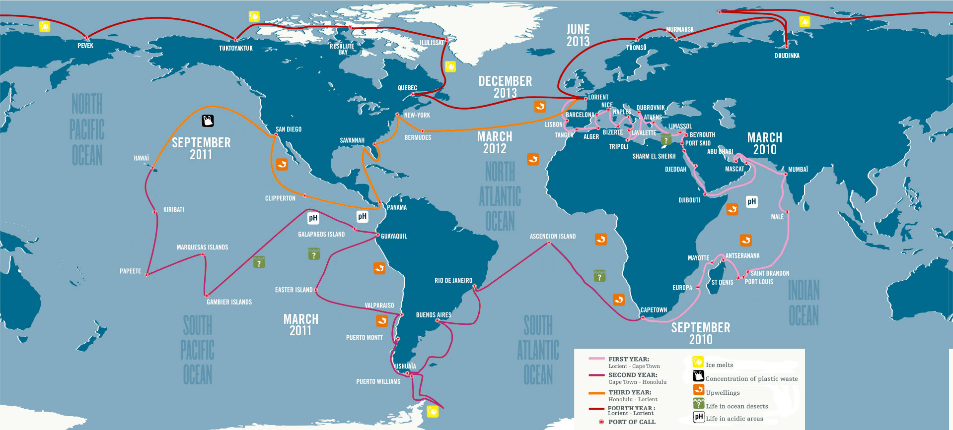
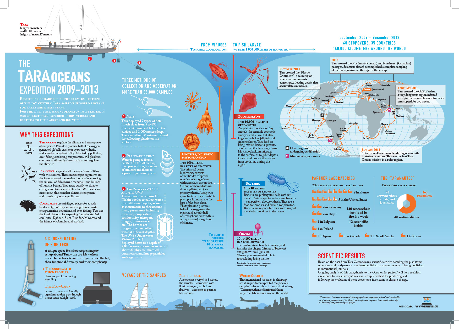
Between 2001 and 2003, Karsenti seemed to be on all fronts. He moved back to Paris to head the Institut Jacques Monod but pursued his EMBL projects. In parallel, he became an advisor to Elisabeth Giacobino, senior researcher at the French Research Ministry. Yet he still had a dream: it was during that period that he hatched the idea of a marine expedition around the world, after reading Charles Darwin’s account of his voyage on board the Beagle. Karsenti’s dream came true with the Tara, a polar schooner that was given a second life as a floating laboratory. In 2009 the Tara Oceans expedition, with Karsenti as scientific director, set sail after a full year of preparation. The ship covered 140,000 kilometers, sailing the Mediterranean, the Red Sea, the Indian, Pacific and Atlantic Oceans, all the way to Antarctica.
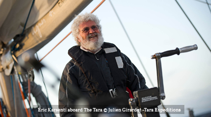 “Tara Oceans was a new adventure for me,” the researcher relates.“At 60, I felt the need to change perspective, to address fascinating but troublesome questions about the past evolution of our planet and its imminent future, in which the oceans are crucial. Not only did life on Earth originate in the oceans, but it still largely depends on them. To improve our understanding of the key role of microscopic marine life, I built up a multidisciplinary team of world-class researchers.
I also wanted to reach out to others, to share the adventure of advancing the scientific understanding of our universe.”
ITINERARY OF THE TARA OCEANS EXPEDITION
© Tara Oceans
THE TARA OCEANS EXPEDITION (2009-2013)
© Tara Oceans
IMAGES OF PLANKTON
PLANKTON CHRONICLES
Credits
Throughout his career, Eric Karsenti has been a pioneer in interdisciplinary approaches to cellular biology. Today, along with Sébastien Colin, he chairs the Morphological Data work group of the Oceanomics project. Providing the scientific follow-up to the 2009 first Tara expedition coordinated by CNRS senior researcher Colomban de Vargas, the group is in charge of collecting, analyzing and archiving the morphological descriptions of the plankton communities sampled during the Tara Oceans expedition. For this researcher who has devoted his professional life to unraveling the secrets of the infinitely small, the adventure continues.
![]() ©2026 CNRS/sagascience
©2026 CNRS/sagascience
Développement : Blueberry Interactive | Legal Notice | Sources & Copyrights | Accessibility | GDPR